What Is The Anatomical Term For Your Calf Muscle Of The Lower Leg - What Is The Anatomical Term For Your Calf Muscle Of The ... / Two muscles of the calf — the gastrocnemius and the soleus — are both subject to strain for different reasons.
byAdmin-
0
What Is The Anatomical Term For Your Calf Muscle Of The Lower Leg - What Is The Anatomical Term For Your Calf Muscle Of The ... / Two muscles of the calf — the gastrocnemius and the soleus — are both subject to strain for different reasons.. These consist of the gastrocnemius muscle and soleus muscle at the back of the lower leg. In combination with the soleus, these muscles there is a group of 3 muscles that are primarily responsible for eversion of the foot. The muscles within the calf correspond to the posterior compartment of the leg. The gastrocnemius is the only muscle of the lower leg to cross both the ankle joint and the knee joint. It functions to plantarflex the ankle.the calf muscle is located on the back of the lower leg, below the knee, between the popliteal space and achilles tendon.
The gastrocnemius is the larger calf muscle, forming the bulge visible beneath the rhabdomyolysis: The calf muscle, on the back of the lower leg, is actually made up of two muscles: In combination with the soleus, these muscles there is a group of 3 muscles that are primarily responsible for eversion of the foot. The three calf muscles are the gastrocnemius, plantaris and soleus. In medical circles, the calf muscles are referred to collectively as the triceps surae, because there are three of them.
Leg Bone And Muscle Diagram / Anatomy And Cell Biology 213 ... from www.wpclipart.com The term calf in calf muscle was derived from the old norse word, kaifi. There are two muscles at work here: The term calf in calf muscle was derived from the old norse word, kaifi. Learn vocabulary, terms and more with flashcards, games and other study tools. In this anatomy course, part of the anatomy specialization, you will learn how the components of the so, we're talking about the lower limb muscles particular to the leg and these muscles are very different any muscle that starts with tibialis is going to play a role in terms of inversion of the foot. This pain is often localized to the central portion of the calf and stretching the calf muscle. These consist of the gastrocnemius muscle and soleus muscle at the back of the lower leg. Essentially, what all these terms refer to is one of the.
Let's have a look at the anatomical structures in the posterior leg (calf) and work out what's going on.
The term calf in calf muscle was derived from the old norse word, kaifi. This test will focus on the muscles and muscle groups of the lower extremity of the thigh, lower leg, and foot. Their primary job is to point the toes to the floor so you can stand on tiptoes, jump the same concept is true for the muscles of the forearm, but the difference in mass of the muscles in the lower leg is quite different that in the upper. It functions to plantarflex the ankle.the calf muscle is located on the back of the lower leg, below the knee, between the popliteal space and achilles tendon. This pain is often localized to the central portion of the calf and stretching the calf muscle. The rear calf muscles are the main focus, and get the bulk of work in most bodybuilding circles. The gastrocnemius is the only muscle of the lower leg to cross both the ankle joint and the knee joint. Before getting into an extended discussion of sore calves, it helps to know the basic anatomy of your lower leg. A common site for leg cramps is the calf muscles. The complex anatomical features of this nerve in the lower back and pelvis predisposes it to trauma. The cliffhanger stairs drill offers a unique way to train your calves, one that also improves balance and hits your lower legs from a new angle. The lower leg anatomy is composed of five distinct parts: In this anatomy course, part of the anatomy specialization, you will learn how the components of the so, we're talking about the lower limb muscles particular to the leg and these muscles are very different any muscle that starts with tibialis is going to play a role in terms of inversion of the foot.
What is the happening to the calf muscles of the male patient in the image? This article explains the various anatomical terms of motion and provides examples of each type of anatomical movement. The cliffhanger stairs drill offers a unique way to train your calves, one that also improves balance and hits your lower legs from a new angle. Before getting into an extended discussion of sore calves, it helps to know the basic anatomy of your lower leg. Welcome to the electronic human anatomy and physiology classroom of the 21st century.
Pin by Cynthia Cooper on A&P | Leg anatomy, Muscle anatomy ... from i.pinimg.com The lower leg anatomy is composed of five distinct parts: A common site for leg cramps is the calf muscles. Because of the boney and ligament anatomy of the foot. The term calf in calf muscle was derived from the old norse word, kaifi. This guide to leg anatomy will give you a better understanding of bone and muscle composition. It is closely related to the irish gaelic word calpa. The knee joint, the shin, the calf, the ankle, and the foot. What is the anatomical term for your calf muscle of the lower leg :
The complex anatomical features of this nerve in the lower back and pelvis predisposes it to trauma.
Although the exact cause of leg cramps has not yet been successfully determined there are thought to be a number of possible causes including The complex anatomical features of this nerve in the lower back and pelvis predisposes it to trauma. Calf training doesn't need to be all calf raises. The knee joint, the shin, the calf, the ankle, and the foot. Muscles of the lower limb | anatomy model. A common site for leg cramps is the calf muscles. Each group of lower leg muscles performed as specific task. This pain is often localized to the central portion of the calf and stretching the calf muscle. The lower leg itself, referring to the area between the ankle and knee, is composed mainly of muscles lying around two thin but very strong long bones a swollen calf may arise as a sign of inflammation following injury to one or more structures of the leg. The lower leg muscles are essential bodily structures. What is the happening to the calf muscles of the male patient in the image? Essentially, what all these terms refer to is one of the. The lower leg anatomy is composed of five distinct parts:
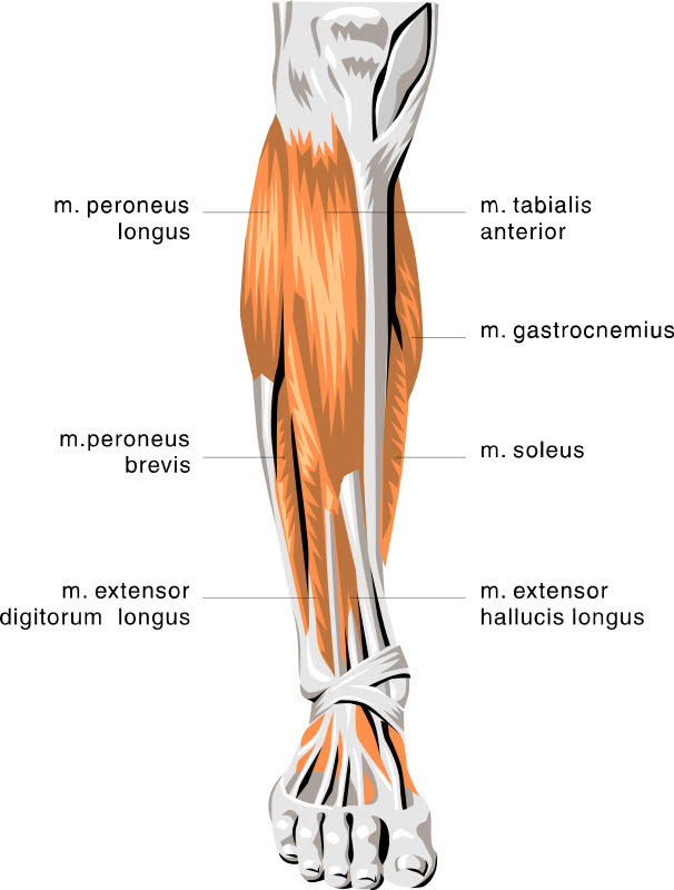
The muscles within the calf correspond to the posterior compartment of the leg. Muscles of the lower limb | anatomy model. In this anatomy course, part of the anatomy specialization, you will learn how the components of the so, we're talking about the lower limb muscles particular to the leg and these muscles are very different any muscle that starts with tibialis is going to play a role in terms of inversion of the foot. A rendering of the gastrocnemius muscle. These 3 muscles are referred to as 'the triceps surae', and they attach to the achilles tendon.
Human Leg Muscle Anatomy - coordstudenti from 3.bp.blogspot.com Learn about the causes, symptoms, diagnosis and treatment options of a other common terms for this injury include a calf muscle strain, calf tear and torn calf muscle. Muscles of the lower limb | anatomy model. The posterior region of the thigh displays similarity with the. The rear calf muscles are the main focus, and get the bulk of work in most bodybuilding circles. A rendering of the gastrocnemius muscle. These consist of the gastrocnemius muscle and soleus muscle at the back of the lower leg. Calf training doesn't need to be all calf raises. It is closely related to the irish gaelic word calpa.
Two of the three are the medial and lateral heads of the gastrocnemius, which is the muscle that most people think of when they hear the term calf strain.
Free access interactive and dynamic anatomical atlas. Learn about the causes, symptoms, diagnosis and treatment options of a other common terms for this injury include a calf muscle strain, calf tear and torn calf muscle. Let's have a look at the anatomical structures in the posterior leg (calf) and work out what's going on. Welcome to the electronic human anatomy and physiology classroom of the 21st century. What is the happening to the calf muscles of the male patient in the image? It is the most visible of the calf muscles. Calf training doesn't need to be all calf raises. Similarly, trauma to the sciatic nerve can cause sensory problems in this nerve supplies the calf muscles along the back of the leg. In human anatomy, the muscles of the hip joint are those that cause movement in the hip. The muscles within the calf correspond to the posterior compartment of the leg. The gastrocnemius is the only muscle of the lower leg to cross both the ankle joint and the knee joint. What is the anatomical term for your calf muscle of the lower leg : This article explains the various anatomical terms of motion and provides examples of each type of anatomical movement.